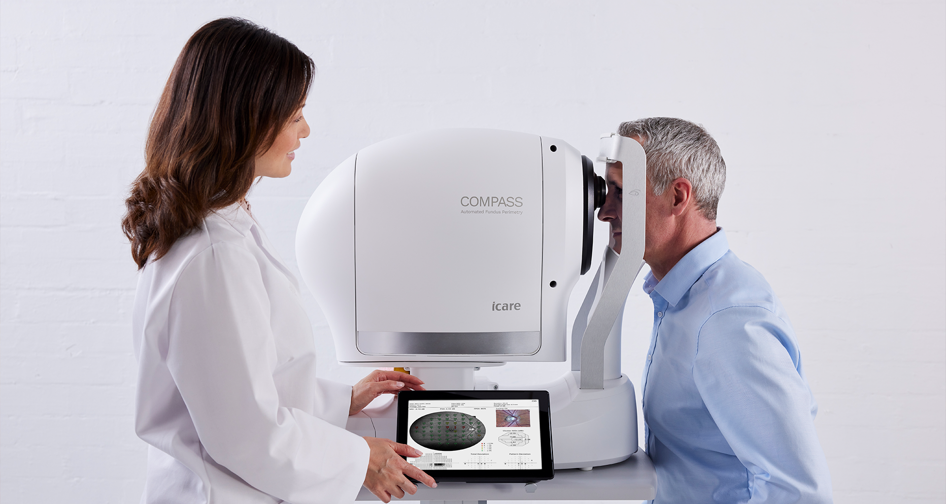
The best of both worlds
The iCare COMPASS now includes ZEST Fast technology, a detailed SPA report and a wider view of the retina.
-
iCare - More than Tonometry
Since 2003 iCare has been known as the original developer of rebound technology. In 2019, automated retinal imaging and perimeters were added to the portfolio increasing iCare's innovative product line.
Find your local iCare specialist -

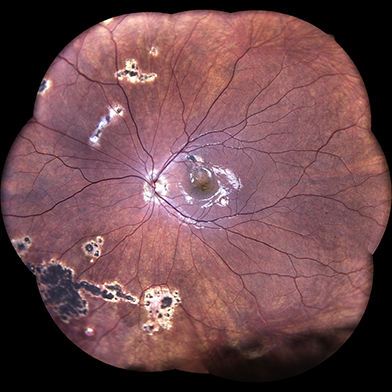
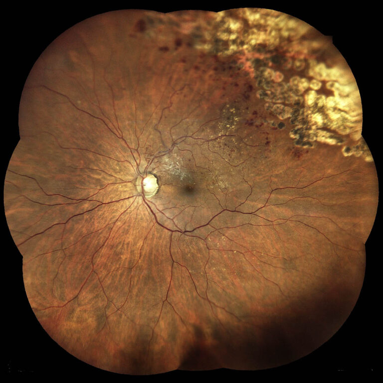
iCare EIDON UWF - TrueColor Confocal Fundus Imaging System
The first TrueColor Confocal imaging device for high resolution ultra-widefield imaging, up to 200° -
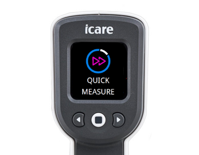
iCare IC200 - Now with Quick Measure!
Introducing the iCare IC200 with Quick Measure. An innovative new feature that makes intraocular pressure (IOP) measurements faster and easier.
iCare Establishes the Next Level of Eye Care
iCare is a trusted partner in ophthalmic diagnostics, offering physicians fast and reliable devices to help diagnosis glaucoma, diabetic retinopathy and macular degeneration. Our product portfolio includes automated TrueColor imaging, perimeters and hand-held rebound tonometers.
We believe in ophthalmic care that is accessible, reliable and effortless. Our mission is to be the leaders in cutting edge products with innovative technology.
iCare is a registered trademark of Icare Finland, Oy. Icare Finland, Oy, Icare USA, Inc., CenterVue S.p.A. and Icare World Australia Pty Ltd. are divisions of Revenio Group representing the iCare brand.
iCare Imaging Systems - with TrueColor Confocal Technology
The iCare advantage of TrueColor captures detail rich images using white LED light to provide superior color fidelity while the confocal imaging blocks scattered light improving image quality. Together they provide enhanced images with greater contrast. iCare imaging systems are fully automated, intuitive and require minimal staff training. Patients will experience reduced waiting times and improved comfort, while office staff gains the benefits of a more efficient workflow.
Imaging devices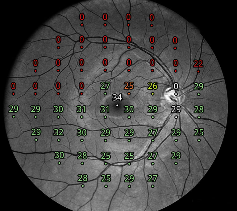
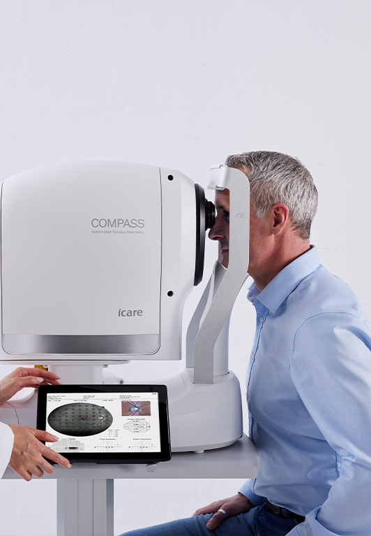
Automated Perimetry with Active Retinal Tracking
iCare's innovative perimeters combine visual field tests, fixation loss correction using real-time retinal tracking and confocal fundus imaging, all in one exam. This ensures an accurate match between function and structure resulting in the reduction of motion artifacts.
Perimeters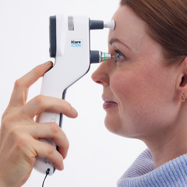
Hand-held Rebound Tonometers
Our patented design allows for quick, reliable IOP measurements. This ergonomic device has an intuitive user interface which ensures easy, safe and accurate operation. Now patients have access to a home tonometer to collect real-world IOP data. This additional data can be used to customize their treatment plans.
TonometersThe Best of Ophthalmic Technology
All iCare’s products are equipped with pioneering technology. iCare imaging systems use a unique confocal TrueColor technology capturing high quality and detail rich images. iCare’s perimeters combined with retinal tracking compensates for eye movements during the test providing more reliable results. iCare tonometers use a patented rebound technology for quick and easy IOP measurements.


-
Where to Buy?
Click below to find your local sales representative.
Find your representative -
Order Probes Today!
Click below to order your sterile probes.
Order Probes